
Connecting Pixels with People
Pathomation offers a complete and scalable Digital Pathology Ecosystem
For Diagnostics, Research, Training & Education
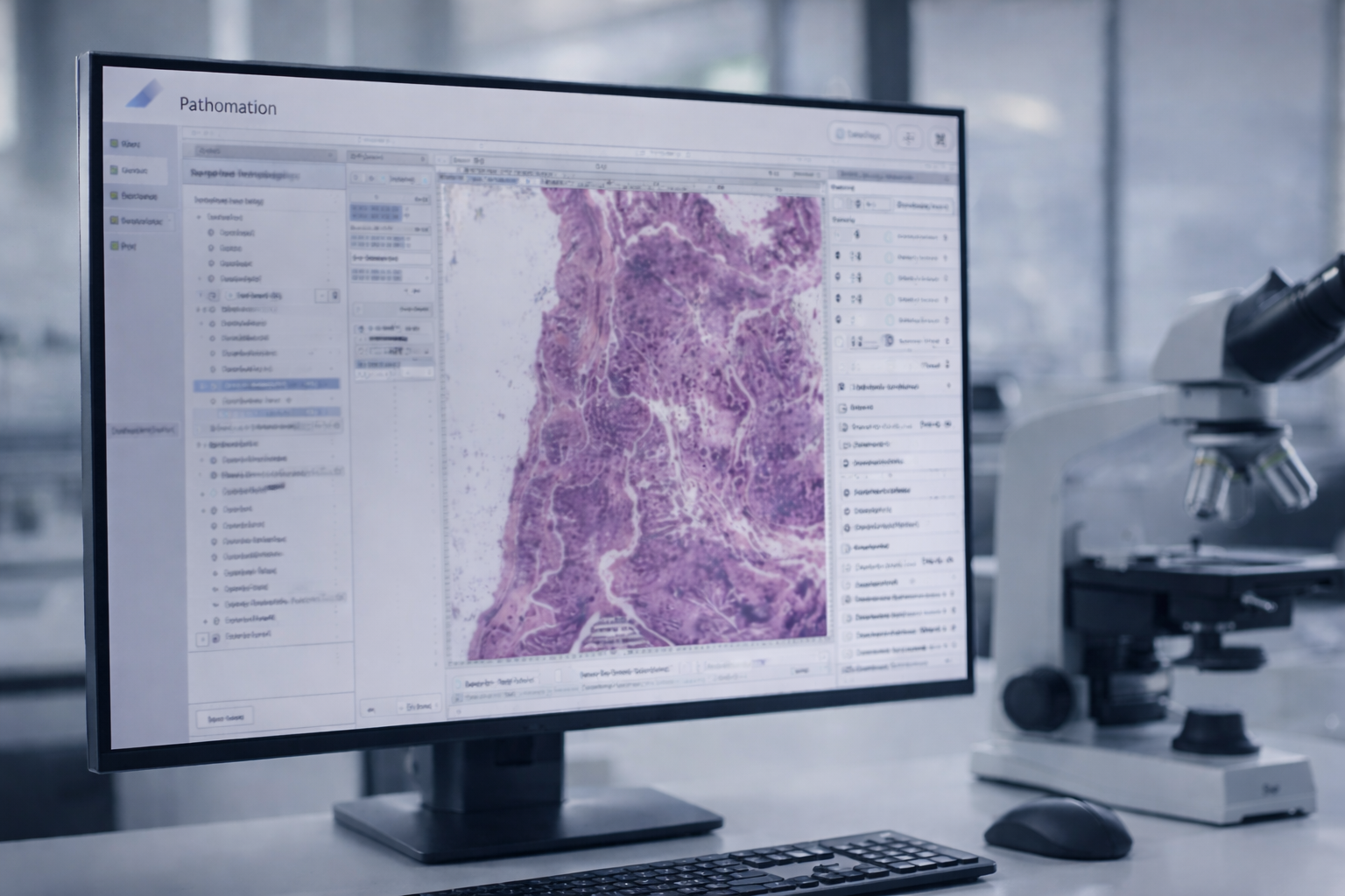
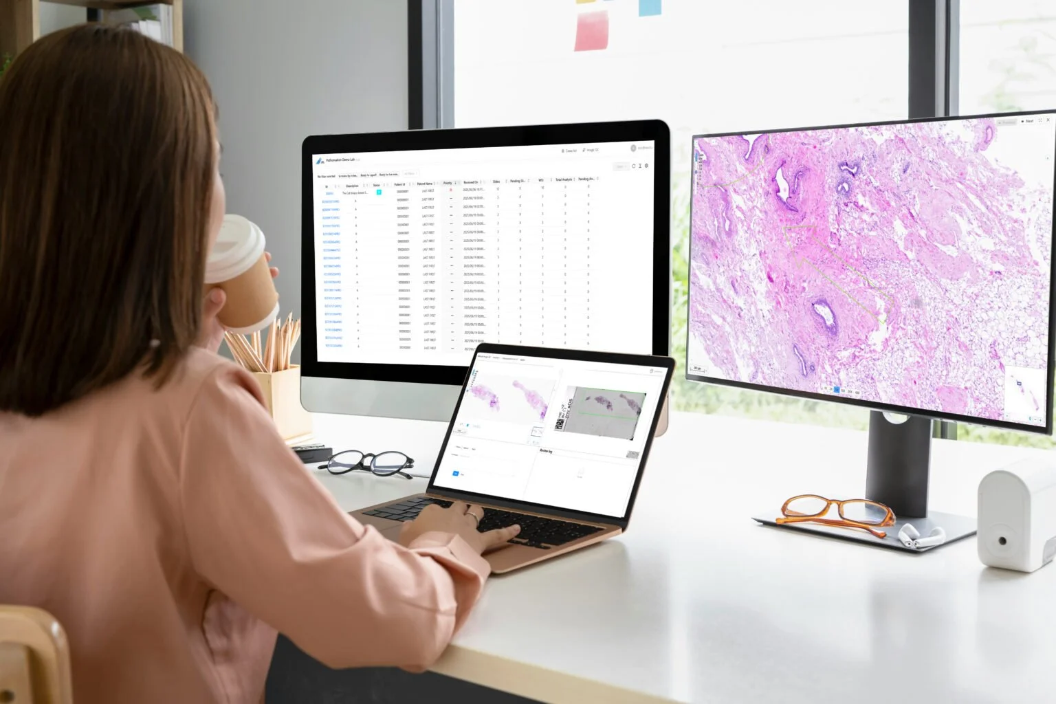
Comprehensive Image Viewing and Management Software for Digital Pathology
DIGITAL PATHOLOGY SOFTWARE FOR EVERY DIGITAL PATHOLOGY FLAVOR
A Modular Software Ecosystem Tailored to Your Needs.
Pathomation adapts to you. Start simple, grow when you're ready. Tailored solutions for every step of your journey.


Ready to Streamline your lab? Get in Touch
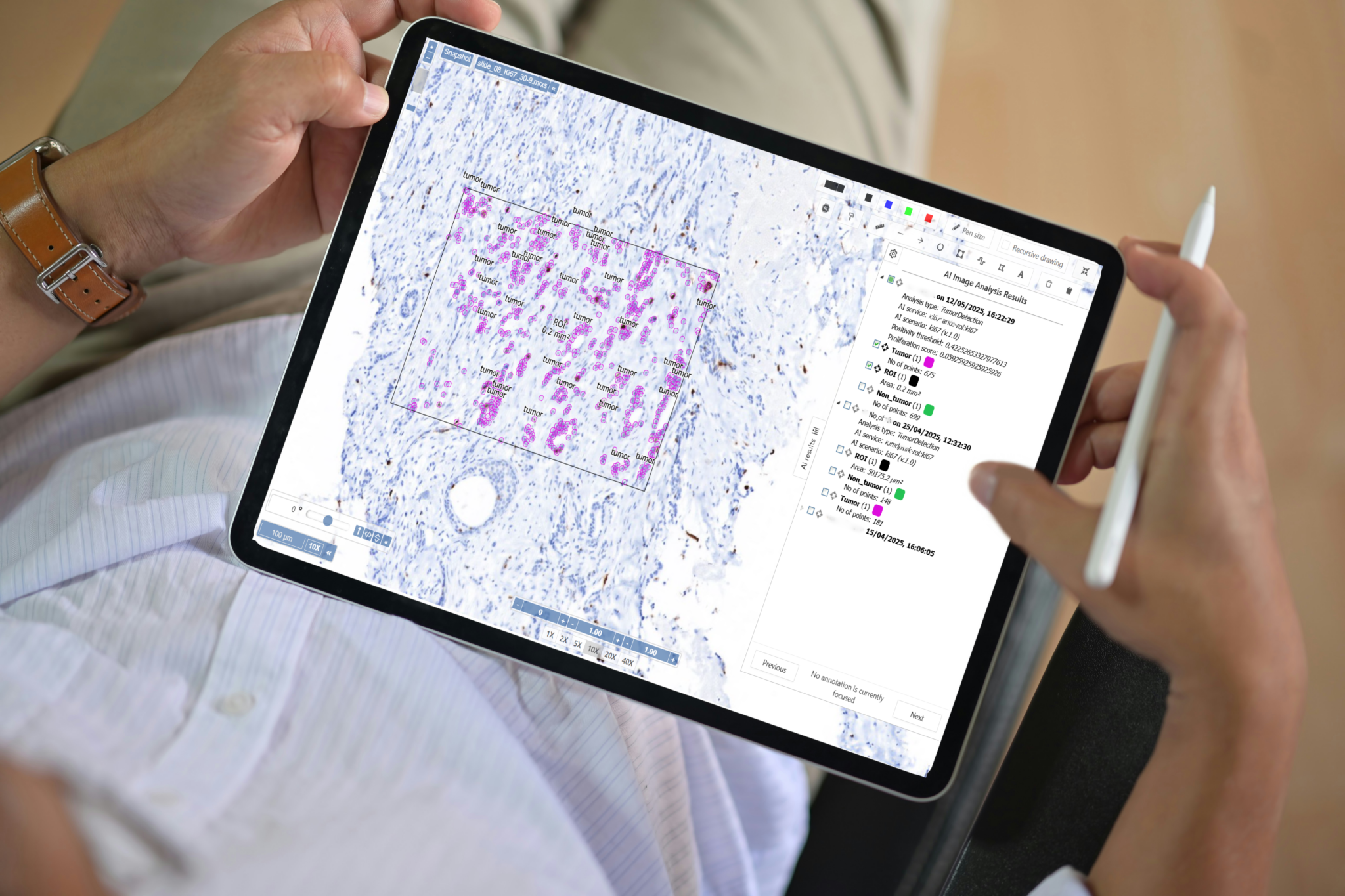
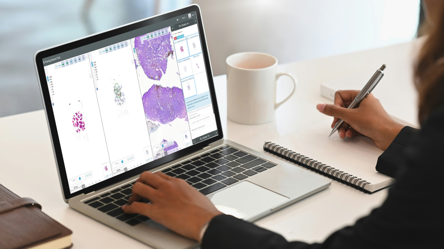
Empower Your Digital Pathology with Seamless AI Integration
Discover how Pathomation’s platform, featuring a built-in AI marketplace, can elevate your research and diagnostics to the next level.
Connect with our experts today and future-proof your lab with smarter, faster, and more accurate pathology workflows.
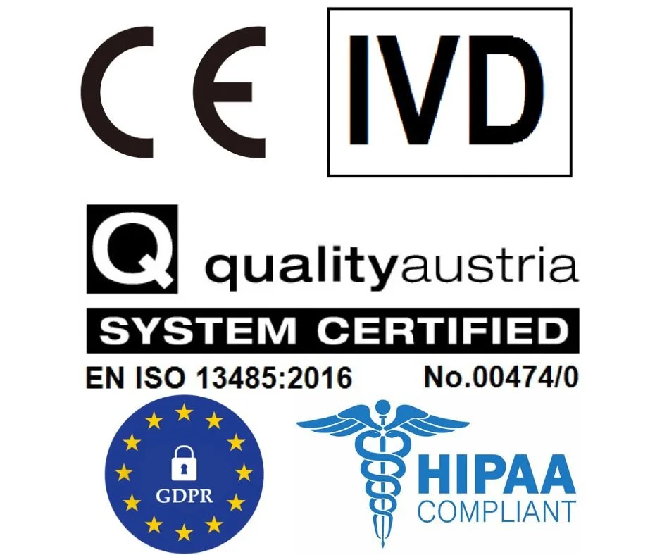
Built on Trust.
Backed by Quality.
At Pathomation, reliability isn’t just an option, it’s the foundation of everything we do. Our software is built to meet the highest standards of security, compliance, and performance, empowering customers in diagnostics, research, and pharma to work with complete confidence.
We follow strict data protection practices, enable traceability, and support regulatory compliance across our solutions. With a track record of long-term partnerships and a proactive approach to quality assurance, we deliver software you can depend on. Today and in the future.
What our customer are saying
Want to stay in the loop?
Get our insights on digital pathology straight to your inbox.
Latest news
My Pathomation update improves user experience and workflow consistency
March 20, 2026
When digital pathology works well, you barely notice the software.
The latest My Pathomation release focuses on exactly that. We focused on improving how complex whole slide image formats are uploaded, handled, and analyzed.
With more consistent DICOM support, guided uploads, and flexible AI workflows for Regions of Interest, daily workflows become smoother and more reliable.
Explore the latest update yourself: https://my.pathomation.com
New Blogpost: Overview of Oncology CDx biomarker educational resources
April 14, 2026
Companion diagnostics have transformed oncology. But their impact depends on one critical factor: How accurately biomarkers are interpreted. Read more…
By Rudy Hovelinck