Evaluation of an online training tool for scoring programmed cell death ligand-1 (PD-L1) diagnostic tests for lung cancer
Bharat Jasani, Gudrun Bänfer, Rebecca Fish, Wim Waelput, Yves Sucaet, Craig Barker, Jessica L Whiteley, Jill Walker, Rudy Hovelinck, Rolf Diezko
Numerous studies indicate that higher tumour programmed cell death ligand-1 (PD-L1) expression is associated with greater response to anti-programmed cell death-1 (PD-1)/PD-L1 immunotherapy in non-small cell lung cancer (NSCLC).
Use of Pathomation’s web-based training tool incorporated into classroom-style training was associated with an overall moderately good level of inter-reader reproducibility at key cut-offs for TC PD-L1 expression testing in NSCLC. Overall, the online training tool offers a means of standardised training for practising pathologists in a clinical setting.
Artificial intelligence assistance significantly improves Gleason grading of prostate biopsies by pathologists
Wouter Bulten, Maschenka Balkenhol, Jean-Joël Awoumou Belinga, Américo Brilhante, Aslı Çakır, Lars Egevad, Martin Eklund, Xavier Farré, Katerina Geronatsiou, Vincent Molinié, Guilherme Pereira, Paromita Roy, Günter Saile, Paulo Salles, Ewout Schaafsma, Joëlle Tschui, Anne-Marie Vos, ISUP Pathology Imagebase Expert Panel, Hester van Boven, Robert Vink, Jeroen van der Laak, Christina Hulsbergen-van der Kaa, Geert Litjens
The pathologists utilized the Pathomation viewer software to assess the biopsies’ grading.
The study explores the impact of integrating artificial intelligence (AI) systems with pathologists’ expertise in Gleason grading of prostate biopsies. It highlights that while AI systems can achieve high performance in grading tasks, their effectiveness within the pathologist’s workflow is still being evaluated. Challenges such as artifacts and rare cancer subtypes can affect AI system assessments, but combining expert opinions with AI feedback can enhance diagnostic accuracy.
The experiment involved pathologists grading biopsies with and without AI assistance, showing that AI significantly improved grading consistency and performance. The study suggests that AI assistance can enhance Gleason grading accuracy and consistency among pathologists, potentially improving prostate cancer diagnosis and patient outcomes.


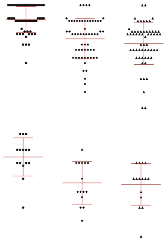
Two Instrument Comparison of Reagents From a US FDA-Approved Assay for the Assessment of Ki-67 in High-Risk Early Breast Cancer
Miglena Komforti, DO,* Erinn Downs-Kelly, DO,* Francisco Sapunar, MD,† Sameera R. Wijayawardana, PhD,‡Aaron M. Gruver, MD, PhD,§ and Sunil S. Badve, MD∥
Toxicologic Pathology Forum: A Roadmap for Building State-of-the-Art Digital Image Data Resources for Toxicologic Pathology in the Pharmaceutical Industry
Digitization of histologic slides brings with it the promise of enhanced toxicologic pathology practice through the increased application of computational methods.
This opinion piece describes the collective experience of building resources for WSI data in the Roche group and elaborates on the challenges encountered and solutions developed with the goal of providing examples of how to build a data resource for digital pathology analytics in the pharmaceutical industry.
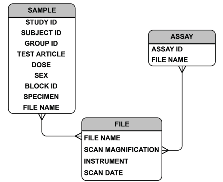
The process of building robust image data resources is challenging and includes considerations of generation, curation, and storage of WSI files, and WSI access including via linked metadata.
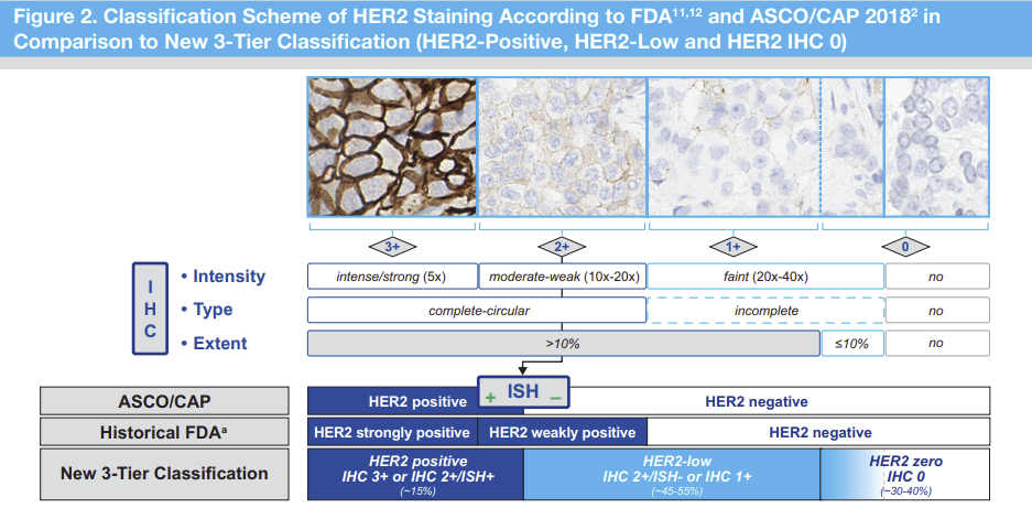
The new HER2 low classification necesssitated retraining of pathologists and assess the accuracy with a proficiciency test
Proficiency Assessment of HER2-Low Breast Cancer Scoring With the Ventana PATHWAY 4B5 and Dako HercepTest HER2 Assays and the Impact of Pathologist Training
Josef Rüschoff, Alexander Penner,Ian O. Ellis, Elizabeth Hale Hammond, Annette Lebeau, Robert Y. Osamura, Fréderique Penault-Llorca, Federico Rojo, Neil Atkey, Andreas H. Scheel, Corrado D´Arrigo, Hans-Ulrich Schildhaus, Akira Moh, Chirag Desai, Giuseppe Viale
This study used the Pathotrainer software to compare Pathologists concordance on HER2 low scoring.
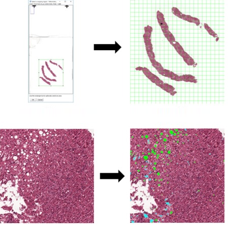
A Novel Automatic Digital Algorithm that Accurately Quantifies Steatosis in NAFLD on Histopathological Whole-Slide Images
Isabelle D Munsterman, Merijn van Erp, Gert Weijers, Carolien Bronkhorst, Chris L de Korte, Joost P H Drenth, Jeroen A W M van der Laak, Eric T T L Tjwa
This paper is an illustration of how PMA.start can be used in a research setting
We developed a digital automated quantification of steatosis on whole-slide images (WSIs) of liver tissue and performed a validation study. Hematoxylin-eosin stained liver tissue slides were digitally scanned, and steatotic areas were manually annotated. We identified thresholds for size and roundness parameters by logistic regression to discriminate steatosis from surrounding liver tissue. The resulting algorithm produces a steatosis proportionate area (SPA; ratio of steatotic area to total tissue area described as percentage). The software can be implemented as a Java plug-in in FIJI, in which digital WSI can be processed automatically using the Pathomation extension.

International consensus guidelines for scoring the histopathological growth patterns of liver metastasis.
“Chapter 1: Introduction” In: Digital Pathology – the Definitive, Complete Reference of Digital Pathology
Liron Pantanowitz, Yves Sucaet, Anil Parwani
An extraordinarily comprehensive and complete book for individuals with anything from minimal knowledge to deep, accomplished experience in Digital Pathology.

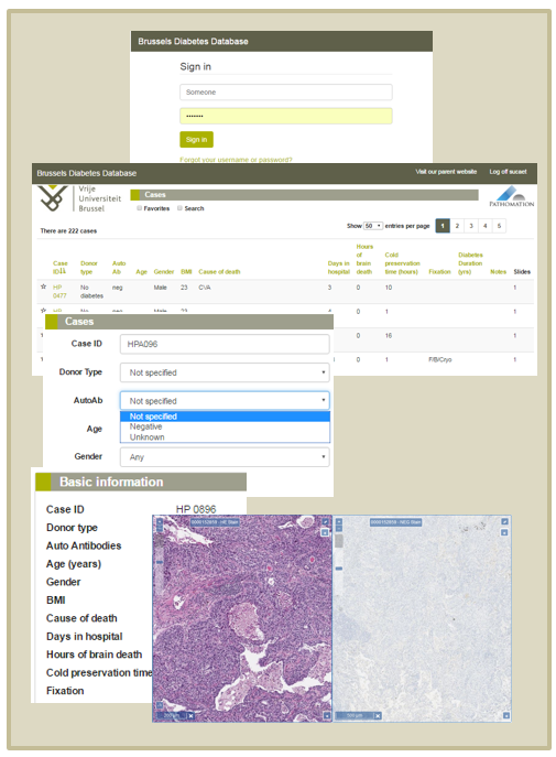
Implementing digital pathology for biobanking
Yves Sucaet
The flexibility of digital pathology hardware and software solutions allows institutions, such as the Brussels Free University, to create bespoke solutions to meet individual biobanking needs
Biobanken gaan digitaal aan de Vrije Universiteit Brussel
Yves Sucaet
Biobanken worden beschouwd als essentieel voor de bevordering van onderzoek en ontwikkeling in de levenswetenschappen. Translationeel biomedisch onderzoek is gebaseerd op grote verzamelingen hoogwaardige proefstukken gecombineerd met grote sets van goed gedocumenteerde gegevens van patiënten en controles. Dergelijke collecties zijn van het allergrootste belang in wetenschappelijke proeven, zowel aangestuurd door onderzoekers als door bedrijven.
Dag microscoop, hallo beeldscherm!
Jacko Duker , Yves Sucaet
Nederland is koploper inzake digitalisatie van medische beeldvormende data. Eigen bedrijven zoals Philips staan hier zelfs wereldwijd om bekend. In dit artikel nemen we u mee naar de pathologie om te zien hoe de opgedane ervaring met digitale technieken het landschap hier in hoog tempo gaat veranderen.
Digital Pathology
Y.Sucaet, W.Waelput
Book published as part of the series: SpringerBriefs in Computer Science
Digital pathology has experienced exponential growth, in terms of its technology and applications, since its inception just over a decade ago. Though it has yet to be approved for primary diagnostics, its values as a teaching tool, facilitator of second opinions and quality assurance reviews, and research are becoming, if not already, undeniable. It also offers the hope of providing pathology consultant and educational services to underserved areas, including regions of the world that could not possibly sustain this level of services otherwise. And this is just the beginning, as its adoption by the also rapidlyemerging fields of medical systems biology and 3D tissue imaging indicate.
This work describes how digital pathology not only has the potential to dramatically impact medical education and the delivery of health care, but also to exert an immensely positive influence worldwide, including in countries and regions that normally fail to benefit from such technological advances.