My Pathomation is a cloud based platform for storing, sharing and annotating images for all scanner file formats.
It comes with advanced features including:
live presentations | slide collections | course & exam building
Scanned microscopy images come in many formats. These large images also need to be shared with many people.
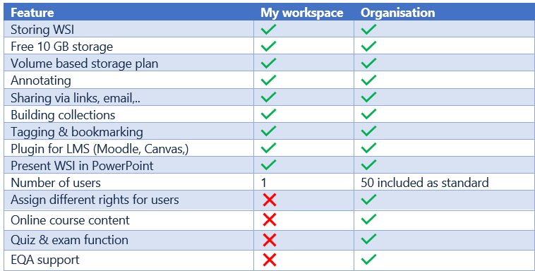
For individuals looking to access and annotate their slide collection anytime, anywhere and from any device – setting up a personal account on My Pathomation is for you.
Whether you’re a teacher sharing virtual slide collections with your students, or a researcher discussing images with international collaborators, setting up an Organisation on My Pathomation can help you
How Does My Pathomation Work?
Register
an individual or organisation account
Upload
your digital pathology slides from your drives to the My Pathomation cloud
Share
slides, slide collections and slide-based content with others privately or publicly
Learn
at your own pace in online courses and exams for students that have text instructions and interactive digital pathology slides
Teach
with live digital slide microscopy in your PowerPoint presentations or classes
With My Pathomation you have one digital microscope that can open all scanners image formats. There is no need to have multiple viewer software packages to open different types of scanner image formats. My Pathomation works entirely via your internet browser and has the added convenience not to require local software installations. Even complex image file formats with Z-stacks or fluorescent images are possible.
Where ever your slides come from, whatever format they are in or where ever your audience resides My Pathomation is the place where all your needs are served. You can upload slides in a drag and drop style to your My Pathomation account. With PMA.start you can connect the My Pathomation cloud storage directly to your local drives and you can manage complex uploads and folder creation at the same time.
Once you have uploaded your slides we have foreseen that you can organise them in different ways to match the logic of your application. You can opt for a folder structure, add patient cases, build collections or design a teaching course. Finally you can add name tags to slides that allow easy searching and organising for large data sets.
Slide organisation is built around:
My Slides – all your slides are here, the view is a classical collapsible file/folder tree, you can group slides in cases
My Collections – the slides that are part of a collection are here, slides can belong to multiple collections, cases can be part of a collection, the view is of images grouped per case/collection
My Slideboxes – this is a collection of collections
My Courses – this is a set of slides or cases you want to use for teaching, you can build a syllabus containing text and slides for student self-paced learning
If you decide one of your collections has scientific or educational value and would benefit the other My Pathomation users you can grant public access to this collection. Similarly you as a user can browse in the public repository for interesting collections and re-use slides to complete your own content.
Sharing and presenting images
A picture says more than 1000 words. But microscopy pictures are huge and difficult to share. Through the My Pathomation platform, you have different options to overcome this difficulty. There is a solution for each situation to allow others to access the images you want to share.
During video conferences or classroom teaching My Pathomation functions like a virtual microscope and your audience can see how you navigate in the images. Whole slide images can be annotated with arrows, text, boxes etc. for efficient and fast navigation.
You can easily create hyperlinks that you can share with colleagues via e-mail or in a chat. Or generate a QR code during a conference presentation as a convenient way for the audience to scan and open the exact same image on their smartphones or tablets.
When people open the image, they will see exactly the same region of interest (ROI) that you are looking at, with the added convenience of still being able to explore the remainder of the virtual slide, too.
Digital pathology user groups
With your pathomation account you can simply work on your own or house the images for your Organisation. You can add other users that belong to your organisation and even create multiple groups.
My Pathomation comes with a set of different user types and rights that reflect the roles you need to run an efficient collaborative project or teach to students. You can define a Manager that will be the administrator of the organisation and has all possible rights. The Editor role is centred on people that work with the actual images and have to develop image content, collaborate with others, present images at conferences or teach students. In contrast a Member is a passive role that can only access images and content without modifying it, typically his will be a student.
Managers and Editors are in charge of adding Members and deciding which content they can see. You can easily create groups and manage to which content the members in the group have access. Adding members can be done by sending a simple e-mail invitation to the people you want to add.
Embedding of images in other content
You can embed the images stored in the My Pathomation cloud in other online solutions and use the universal microscope viewing capacity of My Pathomation.
If your Academic institution uses e-learning platforms like Moodle or Canvas image editing with My Pathomation is built in the software thanks to the ‘Pathomation’ plugin.
Custom website design with WordPress is another application for which we have a plugin. You can learn more about plugins here.
After successfully testing the Pathomation application on our Test Moodle site, we have since deployed it to our live production Moodle site. Much to the delight of our Pathology department. The Pathomation customer service was most helpful and patient in trying to get the application working. They were a pleasure to deal with and very professional. I know the Pathomation application will be a great tool for teaching and the students will benefit immensely from it.
Alan O'Gorman / RCSI Ireland
MyPathomation is an outstanding platform for my needs. It can host my slide collection, make it accessible to outside students and researchers and allow for multiple sorting and organizing of all or part of the collection for in-person or online didactic presentations.
Zev Leifer, PhD. President & CRO / Leifer Institute for Molecular and Digital Pathology
The more we use My Pathomation, the more we like it.
Tyler Anderson / Vitro Molecular Laboratories, USA
I really would like to thank you for this platform, it’s like a dream come true.
This website uses cookies to improve your experience. We'll assume you're ok with this, but you can opt-out if you wish. Cookie settingsACCEPT
Privacy & Cookies Policy
Privacy Overview
This website uses cookies to improve your experience while you navigate through the website. Out of these cookies, the cookies that are categorized as necessary are stored on your browser as they are essential for the working of basic functionalities of the website. We also use third-party cookies that help us analyze and understand how you use this website. These cookies will be stored in your browser only with your consent. You also have the option to opt-out of these cookies. But opting out of some of these cookies may have an effect on your browsing experience.
Necessary cookies are absolutely essential for the website to function properly. This category only includes cookies that ensures basic functionalities and security features of the website. These cookies do not store any personal information.