
Overview of Oncology Companion diagnostic (CDx) biomarker education resources
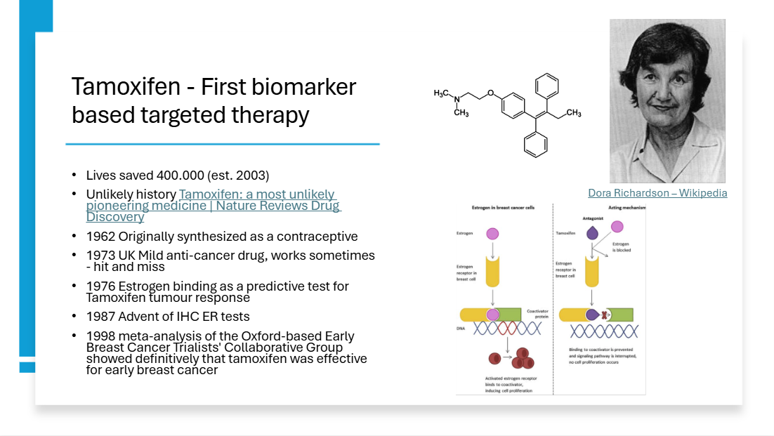
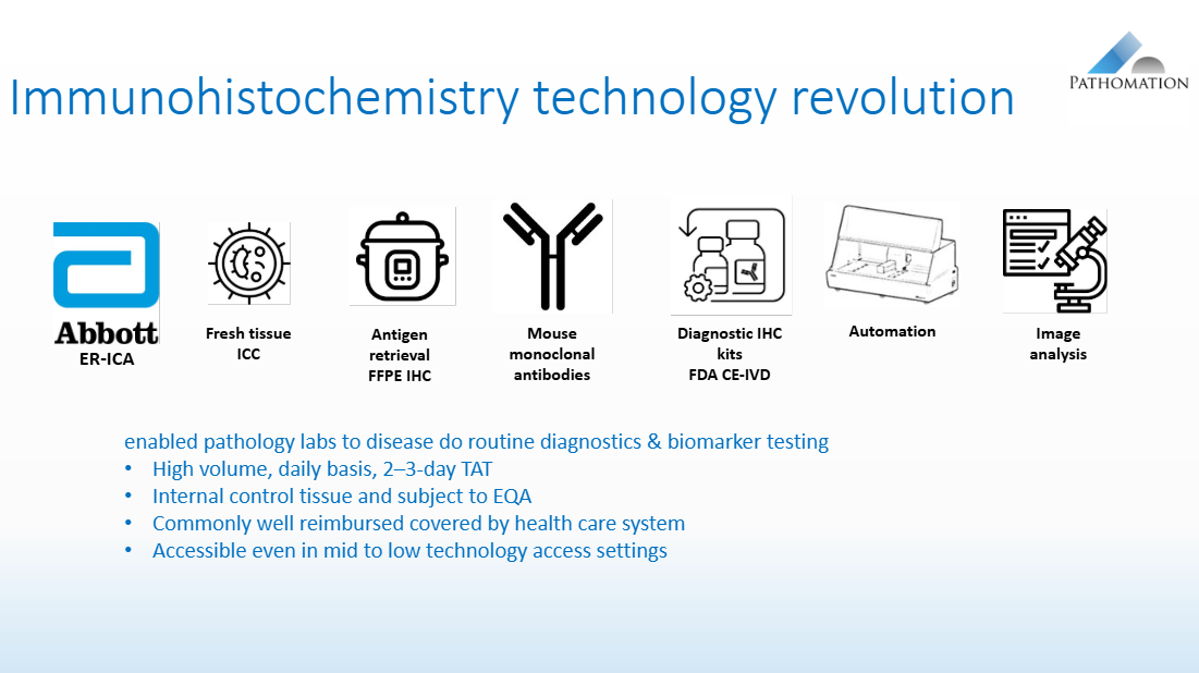
The era of companion diagnostics started with the very first targeted therapy when it was found that Tamoxifen was effective for breast cancer patients overexpressing the Estrogen Receptor. This first biomarker ER gave rise to a revolution in staining techniques that resulted in the highly automated IHC staining methods for formalin fixed paraffin embedded tissue biopsies we use today.
The IHC + targeted therapy model was successfully repeated for a wave of targeted therapies that are now used for treating cancer patients. To name the most well-known biomarker therapy combinations ER/PR – hormone therapy in breast cancer, HER2 – anti HER2 antibody therapies; PD-L1 immune therapy. The evolution of IHC technology enabled the wide adoption of CDx testing that is both reliable, accessible and fast.
Different to simple molecular testing IHC CDx tests require interpretation by a pathologist following a scoring system. These interpretation scoring systems complement the IHC CDx biomarker test and are crucial to make the right therapy selection for the patient. Mistakes in therapy selection can cause direct harm to the patients if they miss out on their most optimal therapy due to a misinterpretation in the scoring and also has a direct societal cost if precious healthcare budget is spent on wrongly prescribed targeted therapies.
In my 30-year experience with CDx biomarkers I have learned that the education of pathologists on biomarkers is often overlooked or if available not fit for purpose. The first reason this is important is to provide training on how to use the biomarker in routine practice so they can reliably report a correct result to their oncologists. The second reason is to make sure that new therapies can reach all of the patient population that will benefit in the shortest possible time frame. Simply put without a trained pathologist a hospital cannot perform the test and complex send out schemes need to be organized which take time to set up. If it takes 3 years for all of the pathologists to perform the CDx test, during that 3-year period a significant number of patients will miss out on their therapy. Generally these will be patients that are less fortunate and do not have the means to travel to an expert center confirming the observation that low socio-economic status may reduce access to testing and that this has important implications for slow uptake of drug prescription.
The pharmaceutical industry has a key role in providing medical education regarding their therapies and companion diagnostic testing since they have all the data including biomarker stained slides generated during clinical trials and can start building diagnostic resources well in advance to drug launches. Professional societies and academic initiatives are slower to generate data and typically lack the resources required to launch global training initiatives.
What matters to pathologists is that the educational resources include access to biomarker stained slides, since the advent of digital pathology it is possible to offer a hands on digital microscope experience and interact with these images via a standard web browser. This has led to a number of e-learning initiatives to support improving biomarker scoring skills.
I wanted to generate a structured, curated list of oncology companion diagnostic biomarker educational resources with digital pathology images to facilitate those looking for more information. This list is not exhaustive but includes all the most relevant resources and a few examples that I personally believe to be very well made. These are my top 11 favorite resources:
-
Link: https://belgian-society-pathology.eu/working-groups/dermato
Content & purpose
Professional working group resources focused on dermatopathology and related biomarkers.
Includes educational documents for members, consensus materials, and presentations on diagnostic and predictive biomarkers.
Target users
Practicing pathologists that are member of the professional society.
Digital pathology component
Interactive digital slide content, PD-L1 stained slides with a focus on the difficulty of interpreting PD-L1 stained slides in the context of melanin pigment in skin samples.
Reference slide sets and knowledge tests are included
-
Link: https://www.claudin182.com/
Dedicated educational platform for the CLDN18.2 companion diagnostic biomarker in gastric/GEJ cancers.
CLDN18.2 is a predictive biomarker used to guide targeted therapy decisions in oncology.
Covers:
Biology and clinical relevance of CLDN18.2
Sample preparation and testing workflows
Stain interpretation and reporting
Expert-led video tutorials
Pathologists, oncologists, and lab professionals involved in CDx testing.
Digital pathology component (strong)
Whole-slide image–like stain galleries
Interactive quizzes for interpretation training
Visual comparison of normal vs tumor staining patterns
Explicit focus on IHC scoring reproducibility
This is one of the best-in-class examples of integrated digital pathology education for a specific biomarker.
-
Link: https://www.msdconnect.sg/therapeutic-areas/biomarkit-overview/biomarker-image-bank-tool/
Content & purpose
Centralized biomarker education platform covering multiple oncology biomarkers.
Country specific, requires registration
Provides:
Overview of biomarker testing strategies
PD-L1, MSI & other companion diagnostic context (therapy linkage)
Image-based learning modules
Target users
Pathologists, oncologists, and diagnostic laboratories.
Digital pathology component (very strong)
Image bank tool with annotated biomarker staining examples
Enables visual benchmarking of:
Positive vs negative cases
Scoring for PD-L1 TPS and CPS cut-offs
Designed to improve interpretation consistency across labs
Key value: cross-biomarker image standardization resource.
-
Link:https://www.idlung.com/home/biomarker.html
Content & purpose
Educational hub focused on all lung cancer biomarkers (e.g., EGFR, ALK, PD-L1).
Includes biomarker biology, testing workflows, and clinical relevance.
Target users
Thoracic oncologists, pathologists, molecular labs.
Digital pathology component
Includes illustrative images and diagrams, sometimes histology/IHC examples.
Less interactive than dedicated image banks but still supports visual learning.
-
Link: https://www.explore-fralpha.com/index.html
Content & purpose
Educational hub focused on all FRalpha biomarker
Includes biomarker biology, a case atlas, and clinical relevance in ovarian cancer.
Target users
Oncologists and pathologists.
Digital pathology component
Case atlas with histology/IHC examples.
Supports self assessment.
-
Link: https://www.myastrazeneca.be/nl/therapeutical-areas/diagnostic/breast-cancer-her2.html
Content & purpose
Educational hub focused on all Breast cancer biomarkers with details on HER2
Representative for local situation in Belgium specific for testing guidelines, drug approvals and reimbursement
Includes biomarker biology, scoring guides, a case atlas, and access to AI assisted scoring.
Target users
Oncologists and pathologists.
Digital pathology component
Case atlas with HER2 IHC examples.
Self assessment for HER2 scoring.
HER2 AI assisted scoring try out.
-
Link: https://patcha.pathomation.com/
Gamification of HER2 scoring.
Short exercise to test your skills in scoring the HER2 stain, the platform returns participants results including time, accuracy and ranking compared to others
Target users
Digital pathology component (advanced)
Region of interest based image recognition based on the well-known captcha test
-
Link: https://www.tilsinbreastcancer.org/tils-pd-l1-training-course/
Content & purpose
Training on:
Tumor-infiltrating lymphocytes (TILs)
PD-L1 scoring across assays
Target users
Breast pathologists, immuno-oncology specialists.
Digital pathology component (very strong)
Access to digitized H&E and PD-L1 slides
Multi-assay comparison (SP142, SP263, 22C3)
Unique for cross-assay concordance training on identical cases.
-
Link: https://pathology-education.agilent.com/en/home.html
Content & purpose
Dako Agilent CDx tests focussed training ecosystem covering PD-L1 (clones 22C3, 28-8) & HER2 Herceptest.
Includes:
E-learning modules
Webinars
Atlas of stains with self assessment possibility
Interpretation guidelines
Assay-specific IFUs
Target users
Pathologists at all experience levels.
Digital pathology component
“Atlas of Stains” with real-case stained tissue images
Training cases with image-based scoring exercises
Excellent for Dako CDx digital training.
A US focussed Deep-dive training for PD-L1 scoring (TPS, CPS) is also available https://pdl122c3-learning.dako.com/us/
-
Link: https://diagnostics.roche.com/us/en/eservice-overview/training-and-education.html
Content & purpose
Ventana CDx tests focussed training ecosystem covering PD-L1, HER2 and other antibodies.
Includes:
E-learning modules
Webinars
Whole slide libraries with self assessment possibility
Interpretation guidelines
Assay-specific IFUs
Target users
Pathologists using Ventana assays and instruments (requires Roche a user account).
Digital pathology component
real-case stained tissue images
-
Link: https://dls.com/biomarker-academy-training/
Content & purpose
On demand global training programs for PD-L1, HER2, MSI, CLDN18.2.
Combines:
Theory
Practical microscopy sessions
Expert-led discussions
Target users
Practicing pathologists and clinical trial labs.
Digital pathology component (very strong)
Delivered using Pathomation digital slide platforms
Includes interactive WSI-based training sessions
Notable for large-scale global training (>6000 pathologists).
Conclusions
The most advanced CDx education tools combine IHC scoring guidance with a whole slide image atlas and self assessments. More advanced topics like common artifacts, how to deal with borderline cases, expected staining results and controls are best handled in live trainings where participants interact in a virtual multi head microscope setting.
Pathomation has proudly contributed to many of the examples above. Building on our 14 years of expertise in the domain our best practice advice is to provide a combination of a publicly accessible knowledge resource complemented with an on-demand training program for pathologists.
New on the horizon is AI assisted learning. Our own experience with integration AI biomarker interpretation in e-learning shows participant engagement and learning progress strongly benefit. The technical challenges to provide such a learning experience are not to be ignored, if users experience delays or have to learn how to use a complicated interface the first encounter with AI algorithms will be disappointing. We all agree first impressions matter.
Learn more about the Pathomation PathoTrainer program: PathoTrainer | The Platform for Biomarker Training, Scoring and CDx Validation. — Pathomation
References
Norris, R.P., Dew, R., Sharp, L. et al. Are there socio-economic inequalities in utilization of predictive biomarker tests and biological and precision therapies for cancer? A systematic review and meta-analysis. BMC Med 18, 282 (2020). https://doi.org/10.1186/s12916-020-01753-0
Li S, He Y, Liu J, Chen K, Yang Y, Tao K, Yang J, Luo K, Ma X. An umbrella review of socioeconomic status and cancer. Nat Commun. 2024 Nov 18;15(1):9993. https://doi.org/10.1038/s41467-024-54444-2