
Pathomation in Peer-Reviewed Research
Pathomation software is used by academic institutions, research laboratories, CROs, and pharmaceutical companies worldwide. Below is a selection of peer-reviewed publications in which Pathomation technology supported digital pathology workflows.

Ming Sound Tsao a, Keith M Kerr b, Mark Kockx c, Mary-Beth Beasley d, Alain C Borczuk e, Johan Botling f, Lukas Bubendorf g, Lucian Chirieac h, Gang Chen i, Teh-Ying Chou j, Jin-Haeng Chung k, Sanja Dacic l, Sylvie Lantuejoul m, Mari Mino-Kenudson n, Andre L Moreira o, Andrew G Nicholson p, Masayuki Noguchi q, Giuseppe Pelosi r, Claudia Poleri s, Prudence A Russell t, Jennifer Sauter u, Erik Thunnissen v, Ignacio Wistuba w, Hui Yu x, Murry W Wynes y, Melania Pintilie z, Yasushi Yatabe aa, Fred R Hirsch
The Blueprint (BP) Programmed Death Ligand 1 (PD-L1) Immunohistochemistry Comparability Project is a pivotal academic/professional society and industrial collaboration to assess the feasibility of harmonizing the clinical use of five independently developed commercial PD-L1 immunohistochemistry assays. The goal of BP phase 2 (BP2) was to validate the results obtained in BP phase 1 by using real-world clinical lung cancer samples. 81 lung cancer cases were selected, the cases included 21 resections, 20 core needle or bronchial biopsy samples, 18 tumor-positive lymph node excision biopsy or resection samples, and 22 cytological cell blocks.The resulting PD-L1 stained slides were scored digitally by 24 experienced pulmonary pathologists (IASLC Pathology Committee members) from 15 countries across five continents.
Digital scoring was performed by accessing these images with use of the PathoTrainer Digital Pathology software. A glass set of slides was used a reference for the results, and both Pearson correlation and Bolt and Altman’s methods demonstrated very high correlation and agreement between the two methods of reading PD-L1 IHC results.
This study was a breakthrough demonstrating for the first time that certain PD-L1 assays could be used interchangeably and at the same time proving that digital slide scoring was reliable across pathologists even in the context of a complicated biomarker.
Pathomation plays a crucial role in enhancing the efficiency and accuracy of pathological evaluations by enabling rapid, reliable slide comparisons without physical transport, thereby accelerating workflows and reducing costs
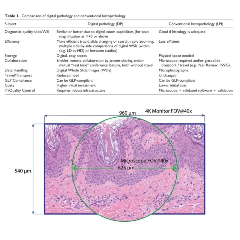
This paper advocates for the integration of digital pathology, specifically whole slide imaging, into toxicologic pathology workflows, including GLP-compliant preclinical safety studies. It highlights the comparable diagnostic accuracy of digital methods to traditional light microscopy, emphasizing advantages such as increased efficiency, remote collaboration, better data management, and compatibility with artificial intelligence. The authors address skepticism about the readiness of digital pathology and present evidence of its validation and regulatory approval, arguing that digital pathology is a reliable, modern alternative for histopathological evaluations in toxicology settings.
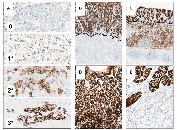
For this study, they used the Pathomation viewer for slide analysis, focusing on the guidelines for evaluating Claudin-18.2 immunohistochemical staining without detailing software or equipment.
The study discusses the evaluation of Claudin-18.2 immunohistochemical staining in gastric and gastroesophageal junction adenocarcinomas to guide targeted therapy. It emphasizes the importance of accurate biomarker evaluation in precision oncology, particularly in the context of limited biopsy material. The authors provide detailed guidelines on tissue preparation, staining procedures, and interpretation of results, highlighting the need for careful assessment to avoid artifacts that could lead to false results. The study also underscores the collaborative efforts of pathologists in optimizing patient selection for therapies like zolbetuximab.
Matteo Fassan, Takeshi Kuwata, Kristina A. Matkowskyj, Christoph Röcken,
Josef Rüschoff
Bharat Jasani, Gudrun Bänfer, Rebecca Fish, Wim Waelput, Yves Sucaet, Craig Barker, Jessica L Whiteley, Jill Walker, Rudy Hovelinck, Rolf Diezko
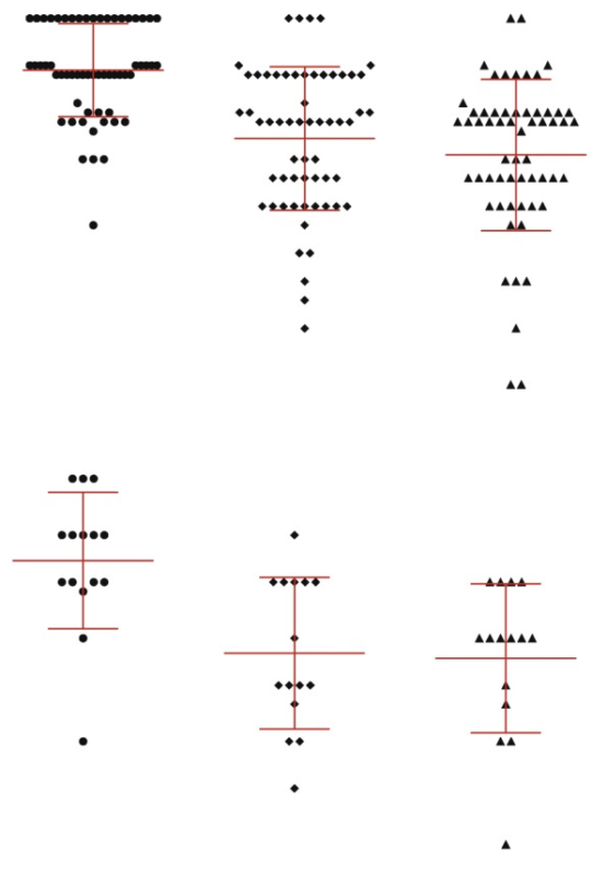
Numerous studies indicate that higher tumour programmed cell death ligand-1 (PD-L1) expression is associated with greater response to anti-programmed cell death-1 (PD-1)/PD-L1 immunotherapy in non-small cell lung cancer (NSCLC).
Use of Pathomation's web-based training tool incorporated into classroom-style training was associated with an overall moderately good level of inter-reader reproducibility at key cut-offs for TC PD-L1 expression testing in NSCLC. Overall, the online training tool offers a means of standardised training for practising pathologists in a clinical setting.
Wouter Bulten, Maschenka Balkenhol, Jean-Joël Awoumou Belinga, Américo Brilhante, Aslı Çakır, Lars Egevad, Martin Eklund, Xavier Farré, Katerina Geronatsiou, Vincent Molinié, Guilherme Pereira, Paromita Roy, Günter Saile, Paulo Salles, Ewout Schaafsma, Joëlle Tschui, Anne-Marie Vos, ISUP Pathology Imagebase Expert Panel, Hester van Boven, Robert Vink, Jeroen van der Laak, Christina Hulsbergen-van der Kaa, Geert Litjens
The pathologists utilized the Pathomation viewer software to assess the biopsies' grading.
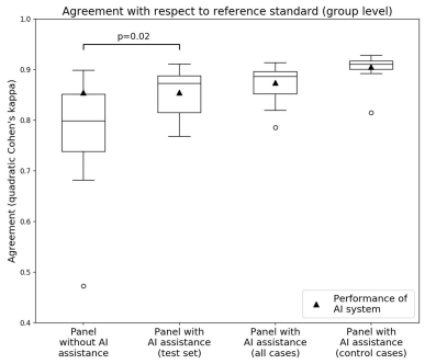
The study explores the impact of integrating artificial intelligence (AI) systems with pathologists' expertise in Gleason grading of prostate biopsies. It highlights that while AI systems can achieve high performance in grading tasks, their effectiveness within the pathologist's workflow is still being evaluated. Challenges such as artifacts and rare cancer subtypes can affect AI system assessments, but combining expert opinions with AI feedback can enhance diagnostic accuracy.
The experiment involved pathologists grading biopsies with and without AI assistance, showing that AI significantly improved grading consistency and performance. The study suggests that AI assistance can enhance Gleason grading accuracy and consistency among pathologists, potentially improving prostate cancer diagnosis and patient outcomes.
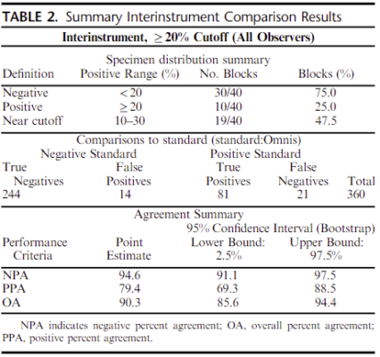
Miglena Komforti, DO,* Erinn Downs-Kelly, DO,* Francisco Sapunar, MD,† Sameera R. Wijayawardana, PhD,‡Aaron M. Gruver, MD, PhD,§ and Sunil S. Badve, MD∥
The objective of this study was to measure concordance of results obtained from the US Food and Drug Administration–approved Ki-67 immunohistochemistry MIB-1 pharmDx assay performed on the Dako Omnis automated staining instrument (Omnis) versus results produced from the assay reagents applied using an optimized protocol on the more widely available Autostainer Link 48 (ASL48) platform. Evaluations were performed utilizing whole-slide images captured using a PANNORAMIC 250 scanner (3DHISTECH, Budapest, Hungary) at ×40 magnification and analysed utilizing the PathoTrainer platform.

Xing-Yue Ge, Juergen Funk, Tom Albrecht, Merima Birkhimer, Moritz Gilsdorf, Matthew Hayes, Fangyao Hu, Pierre Maliver, Mark McCreary, Trung Nguyen, Fernando Romero-Palomo, Shanon Seger, Reina N. Fuji, Vanessa Schumacher, Ruth Sullivan
Digitization of histologic slides brings with it the promise of enhanced toxicologic pathology practice through the increased application of computational methods.
This opinion piece describes the collective experience of building resources for WSI data in the Roche group and elaborates on the challenges encountered and solutions developed with the goal of providing examples of how to build a data resource for digital pathology analytics in the pharmaceutical industry.
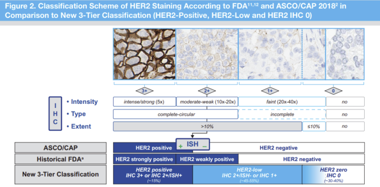
Josef Rüschoff, Alexander Penner,Ian O. Ellis, Elizabeth Hale Hammond, Annette Lebeau, Robert Y. Osamura, Fréderique Penault-Llorca, Federico Rojo, Neil Atkey, Andreas H. Scheel, Corrado D´Arrigo, Hans-Ulrich Schildhaus, Akira Moh, Chirag Desai, Giuseppe Viale
This study used the Pathotrainer software to compare Pathologists concordance on HER2 low scoring.
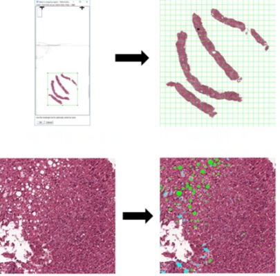
Isabelle D Munsterman, Merijn van Erp, Gert Weijers, Carolien Bronkhorst, Chris L de Korte, Joost P H Drenth, Jeroen A W M van der Laak, Eric T T L Tjwa
This paper is an illustration of how PMA.start can be used in a research setting
We developed a digital automated quantification of steatosis on whole-slide images (WSIs) of liver tissue and performed a validation study. Hematoxylin-eosin stained liver tissue slides were digitally scanned, and steatotic areas were manually annotated. We identified thresholds for size and roundness parameters by logistic regression to discriminate steatosis from surrounding liver tissue. The resulting algorithm produces a steatosis proportionate area (SPA; ratio of steatotic area to total tissue area described as percentage). The software can be implemented as a Java plug-in in FIJI, in which digital WSI can be processed automatically using the Pathomation extension.
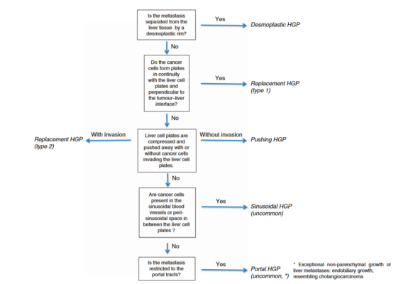
Pieter-Jan van Dam, Eric P van der Stok, Laure-Anne Teuwen, Gert G Van den Eynden, Martin Illemann, Sophia Frentzas, Ali W Majeed, Rikke L Eefsen, Robert RJ Coebergh van den Braak, Anthoula Lazaris, Maria Celia Fernandez, Boris Galjart, Ole Didrik Laerum, Roni Rayes, Dirk J Grunhagen, Michelle Van de paer, Yves Sucaet, Hardeep Singh Mudhar, Michael Schvimer, Hanna Nystro, Mark Kockx, Nigel C Bird, Fernando Vidal-Vanaclocha, Peter Metrakos, Eve Simoneau, Cornelis Verhoef, Luc Y Dirix, Steven Van Laere, Zu-hua Gao, Pnina Brodt, Andrew R Reynolds and Peter B Vermeulen
Pathomation's viewing software was used for the assessment and scoring of histopathological growth patterns (HGPs) of liver metastases.
This publication outlines international guidelines for scoring histopathological growth patterns (HGPs) of liver metastasis, focusing on desmoplastic, pushing, and replacement types. Developed and validated by an international team, these guidelines showed strong scoring agreement.
Desmoplastic HGPs correlate with better survival after liver metastasis surgery compared to other types. The guidelines aim to standardize HGP scoring for better prognosis and personalized treatment, noting desmoplastic HGPs respond better to therapies like bevacizumab due to their angiogenesis reliance. Highlighting HGPs' role in predicting outcomes and guiding treatment, promoting their use as biomarkers in personalized medicine, and enhancing assessment reliability for tailored liver metastasis treatments.
An extraordinarily comprehensive and complete book for individuals with anything from minimal knowledge to deep, accomplished experience in Digital Pathology.
Liron Pantanowitz, Yves Sucaet, Anil Parwani
Yves Sucaet
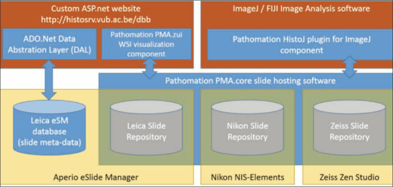
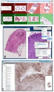
The flexibility of digital pathology hardware and software solutions allows institutions, such as the Brussels Free University, to create bespoke solutions to meet individual biobanking needs
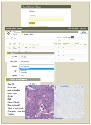
Biobanken worden beschouwd als essentieel voor de bevordering van onderzoek en ontwikkeling in de levenswetenschappen. Translationeel biomedisch onderzoek is gebaseerd op grote verzamelingen hoogwaardige proefstukken gecombineerd met grote sets van goed gedocumenteerde gegevens van patiënten en controles. Dergelijke collecties zijn van het allergrootste belang in wetenschappelijke proeven, zowel aangestuurd door onderzoekers als door bedrijven.
Yves Sucaet
Nederland is koploper inzake digitalisatie van medische beeldvormende data. Eigen bedrijven zoals Philips staan hier zelfs wereldwijd om bekend. In dit artikel nemen we u mee naar de pathologie om te zien hoe de opgedane ervaring met digitale technieken het landschap hier in hoog tempo gaat veranderen.
Jacko Duker , Yves Sucaet
Y.Sucaet, W.Waelput
Book published as part of the series: SpringerBriefs in Computer Science
Digital pathology has experienced exponential growth, in terms of its technology and applications, since its inception just over a decade ago. Though it has yet to be approved for primary diagnostics, its values as a teaching tool, facilitator of second opinions and quality assurance reviews, and research are becoming, if not already, undeniable. It also offers the hope of providing pathology consultant and educational services to underserved areas, including regions of the world that could not possibly sustain this level of services otherwise. And this is just the beginning, as its adoption by the also rapidlyemerging fields of medical systems biology and 3D tissue imaging indicate.
This work describes how digital pathology not only has the potential to dramatically impact medical education and the delivery of health care, but also to exert an immensely positive influence worldwide, including in countries and regions that normally fail to benefit from such technological advances.